General practice is often where complex spinal and neurovascular disease first becomes clinically clear. A patient may have longstanding back or neck pain, then return with worsening gait, declining hand function or new neurological signs that raise concern for degenerative cervical myelopathy. Another may present after incidental imaging has identified a vascular lesion, leaving you to decide whether the finding is significant, how quickly specialist input is needed, and which service is best placed to assess it.
Why an academic referral destination matters
GPs play a key role in reducing avoidable diagnostic delay. Referral decisions are shaped by factors such as symptom burden, neurological findings and functional limitation. Sometimes, a striking scan may sit alongside relatively preserved function, while a modest report may accompany clear deterioration in mobility, independence or work capacity.
An academic referral destination offers a practical advantage in these cases because assessment is not limited to a single procedural opinion. Complex presentations may need subspecialist review, multidisciplinary input and consideration of several management pathways before the most appropriate plan becomes clear. Depending on the diagnosis, that may include surveillance, non-surgical care, open surgery, endovascular intervention, radiosurgery or staged reconstruction.
This model is particularly valuable when the clinical picture is evolving, the diagnosis is high acuity, or the consequences of delay are significant. In patients with suspected cervical myelopathy, complex spinal stenosis, adult deformity, cerebrovascular disease or brain tumour, an academic service can support earlier diagnostic clarification, more precise triage and treatment planning informed by current evidence and cross-disciplinary review.
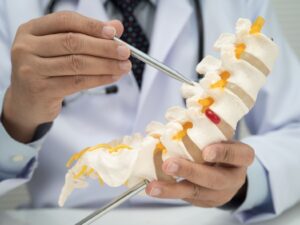
When to refer complex spinal conditions
Most patients with spinal stenosis or degenerative disc disease can be managed conservatively at first, but referral should be considered earlier when the pattern suggests neurological compromise, structural complexity or declining function.
A lower threshold for referral is reasonable when you see:
- progressive motor deficit
- recurrent falls or worsening gait
- hand dysfunction suggestive of cervical myelopathy
- severe neurogenic claudication
- markedly reduced walking tolerance
- bowel or bladder disturbance
- saddle symptoms
- progressive deformity or suspected instability
- recurrent or worsening symptoms after prior spinal surgery
| Suspected condition | Look for | Notes |
| Degenerative cervical myelopathy |
| These features should not be dismissed as routine degenerative change. |
| Clinically significant lumbar stenosis |
| These features are usually more helpful than pain severity alone when deciding whether to escalate referral. |
| Adult spinal deformity |
| Structural complexity, overall alignment and patient-reported disability all affect management decisions. |
When to refer neurovascular conditions
Known or suspected intracranial aneurysms, arteriovenous malformations, arteriovenous fistulas and cerebral cavernous malformations are best referred when the lesion needs more than a descriptive imaging report.
The decision to refer is shaped by factors such as the lesion’s size, location, morphology, symptoms, prior haemorrhage and the patient’s broader risk profile.
A lower threshold for referral is reasonable when you see:
- sudden severe headache or sentinel headache
- new focal neurological deficit or cranial neuropathy
- seizure in the setting of a vascular lesion
- previous intracranial haemorrhage
- known or suspected aneurysm, AVM, AV fistula or cavernous malformation
- progressive neurological symptoms despite apparently small or incidental imaging findings
- pregnancy planning or pregnancy in a patient with a known vascular malformation where multidisciplinary advice may be needed
| Suspected condition | Look for | Notes |
| Unruptured intracranial aneurysm |
| These features can materially change rupture risk and the need for specialist review. |
| Brain arteriovenous malformation |
| Surveillance, radiosurgery, endovascular treatment or surgery may be considered. Management decisions are often multimodal. |
| Cerebral cavernous malformation |
| Symptomatic lesions are managed according to presentation, anatomy and risk. Microsurgery or radiosurgery may be considered. |
Brain tumours and cranial pathology
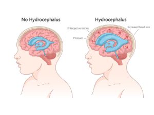
Known or suspected brain tumours and other cranial lesions are best referred when the presentation suggests more than an isolated headache or non-specific neurological complaint.
The decision to refer is shaped by the symptom pattern, pace of progression, focal neurological findings, seizure history, cognitive or behavioural change, functional decline and any imaging abnormality that needs neurosurgical or neuro-oncology input.
A lower threshold for referral is reasonable when you see:
- headache plus seizure, cognitive change or focal neurological symptoms
- first seizure in an adult
- progressive focal weakness, speech disturbance or visual change
- new personality or behavioural change
- unexplained nausea or vomiting with other neurological features
- progressive loss of central neurological function
- declining cognition, independence or work capacity without another clear cause
- imaging showing a cranial lesion that needs specialist assessment or management planning.
| Suspected condition | Look for | Notes |
| Brain tumour or other intracranial mass lesion |
| Symptom combinations are more concerning than headache alone and should prompt earlier investigation or referral. |
| High-grade or progressive cranial pathology |
| Pace of progression helps guide urgency. Progressive loss of central neurological function warrants prompt imaging and specialist assessment. |
| Incidental or newly identified cranial lesion on imaging |
| Management may depend on neurosurgical, neuropathological or neuro-oncology input. |
What to include in the referral
A concise summary of progression, examination findings and the reason for referral is usually more useful than broad statements of concern. Functional detail gives context to imaging and helps the receiving team judge acuity more accurately.
| A meaningful referral aids triage | |
| Referral objective |
|
| Clinical summary |
|
| Examination findings |
|
| Functional impact |
|
| Relevant history |
|
| Imaging |
|
Conclusion
Timely referral can make a meaningful difference in complex spinal, neurovascular and cranial presentations, particularly when symptoms are progressive, neurological function is changing, or the diagnosis is likely to require more than a routine specialist opinion.
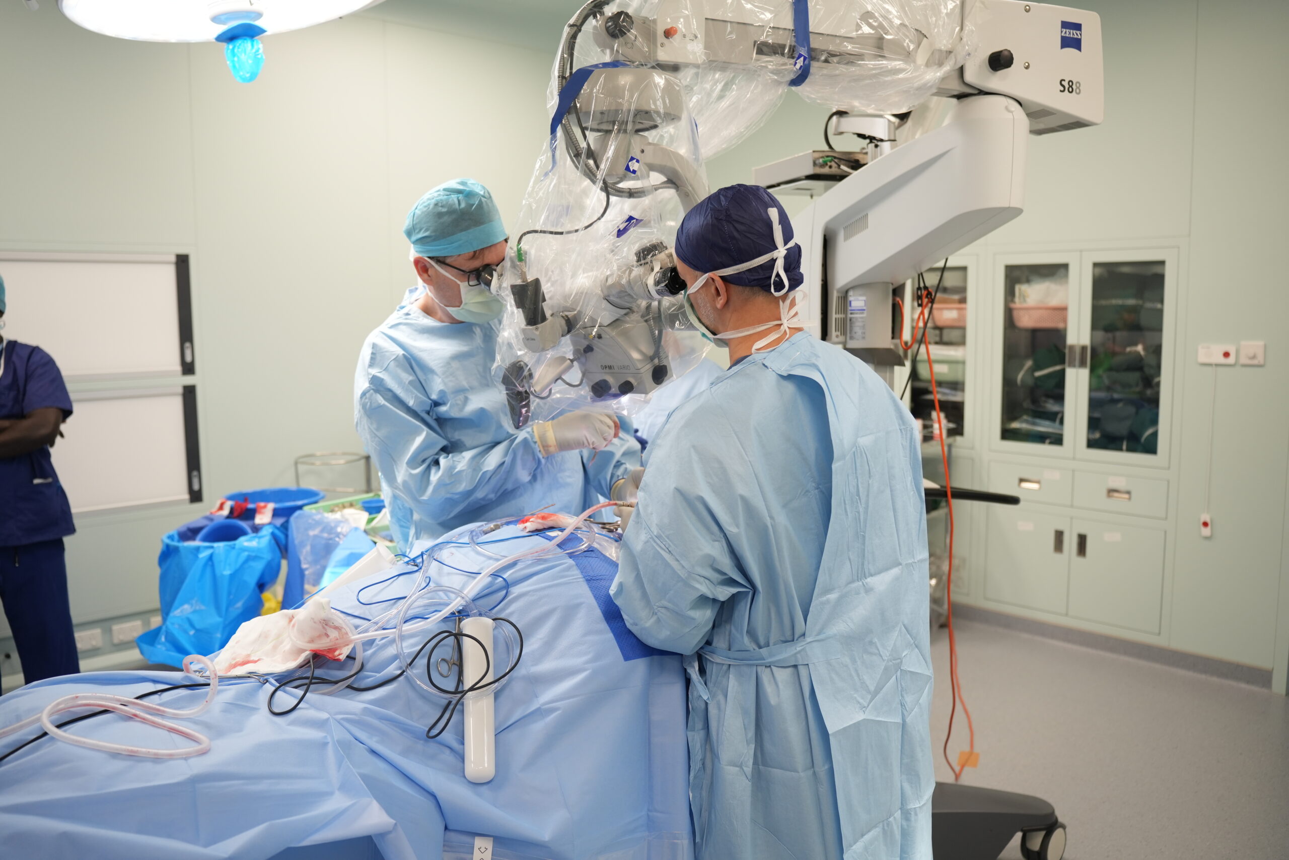
At Macquarie Neurosurgery and Spine, your patient can be assessed within an academic neurosurgical service with subspecialty expertise across spine, neurovascular and tumour care, supported by multidisciplinary review, research-informed planning and advanced technology at Macquarie University Hospital.
Disclaimer
This information is intended for healthcare professionals.
References
- Inzerillo S, Jagtiani P, Jones S. Optimising early detection of degenerative cervical myelopathy: a systematic review of quantitative screening tools for primary care. BMJ Neurology Open. 2025;7:e000913. https://doi.org/10.1136/bmjno-2024-000913, [Accessed 27 March 2026]
- Anderson D, Beard D, Rannou F et al.
Clinical assessment and management of lumbar spinal stenosis: clinical dilemmas and considerations for surgical referral
The Lancet Rheumatology, 2024; 6, e727-e732, https://www.thelancet.com/journals/lanrhe/article/PIIS2665-9913%2824%2900028-6/abstract, [Accessed 27 March 2026] - Diebo, B. G., Shah, N. V., Boachie-Adjei, O., Zhu, F., Rothenfluh, D. A., Paulino, C. B., Schwab, F. J., & Lafage, V. (2019). Adult spinal deformity. The Lancet, 394(10193), 160–172. https://doi.org/10.1016/S0140-6736(19)31125-0, [Accessed 27 March 2026]
- Ogilvy, C. S. (2025). Unruptured intracranial aneurysms. The New England Journal of Medicine, 392(23), 2357–2366. https://doi.org/10.1056/NEJMcp2409371, [Accessed 27 March 2026]
- Ogilvy, C. S. (2025). Unruptured intracranial aneurysms. The New England Journal of Medicine, 392(23), 2357–2366. https://doi.org/10.1056/NEJMcp2409371, [Accessed 27 March 2026]
- Han H, Chen Y, Ma L, et al. Interventional Treatment vs Conservative Management of Unruptured Brain Arteriovenous Malformations. JAMA Netw Open. 2025;8(11):e2543408. doi:10.1001/jamanetworkopen.2025.43408, https://jamanetwork.com/journals/jamanetworkopen/fullarticle/2841324, [Accessed 27 March 2026]
- Rinkel, G. J. E. (2024). Cerebral cavernous malformations: To operate or not? The Lancet Neurology, 23(6), 546–547. https://doi.org/10.1016/S1474-4422(24)00161-3, [Accessed 27 March 2026]
- Weller, M., Preusser, M., Howng, S.-L., Bromberg, J. E. C., Hofer, S., Marosi, C., & van den Bent, M. J. (2023). Primary brain tumours in adults. The Lancet, 402(10406), 1564–1579. https://doi.org/10.1016/S0140-6736(23)01054-1, [Accessed 27 March 2026]
- Booth, T. C., Summers, D., Shah, J., Bulbeck, H., Grant, R., & et al. (2025). Earlier diagnosis of patients with brain tumour. Practical Neurology, 25(6), 504–506. https://doi.org/10.1136/pn-2025-004768, [Accessed 27 March 2026]
- Schaff LR, Mellinghoff IK. Glioblastoma and Other Primary Brain Malignancies in Adults: A Review. JAMA. 2023;329(7):574–587. doi:10.1001/jama.2023.0023, https://jamanetwork.com/journals/jama/article-abstract/2801673, [Accessed 27 March 2026]