
Positional Plagiocephaly
Plagiocephaly is also commonly referred to as “flat head syndrome”. It frequently occurs in babies, where a flat area appears on one side of their head causing an asymmetry of the head shape.
To accommodate for brain, skull and head growth, babies have open cranial sutures and soft spots, called fontanelle. This makes the babies’ skull flexible and malleable. Therefore, a baby’s soft skull can flatten from prolonged pressure in one position. Positional Plagiocephaly occurs when all sutures and fontanelles remain open and does not affect brain development.
Parents can often prevent or treat it with activities like supervised tummy time and by varying the baby’s position during sleep and play. Another common reason for positional plagiocephaly is torticollis, where neck movement is restricted or fixated to certain positions.
Treatment often includes allied health specialists including physiotherapy. In severe cases helmet therapy may also be beneficial.
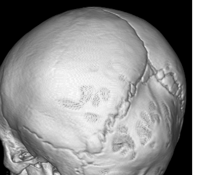
Elements of Craniosynostosis?
Sutures:
A baby’s skull has multiple joints, or sutures, which are typically flexible to allow for brain growth and passage through the birth canal.
Premature closure:
In craniosynostosis, these sutures fuse and become rigid too early.
Skull shape:
As a result the skull grows in an abnormal shape to compensate for the closed suture. Skull appearances may be long, narrow, wide, or uneven.
Symptoms
Symptoms may include the following but are not limited to:
- An unusually shaped head, which may be long and narrow, wider than usual, or uneven.
- A raised ridge or bump along the closed suture.
- A bulging fontanelle (soft spot).
- Lethargy, irritability, difficulties with sleep or feeding.
- developmental delays.
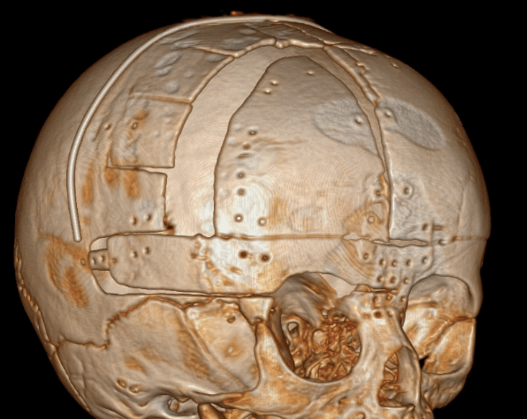
Treatment
Treatment especially in multiple craniosynostosis may prevent high pressure inside the skull that can lead to developmental delay and other neurological deficits. For single craniosynostosis treatment is often but not always for cosmetic reasons. Surgical intervention can help create room for the brain to grow and correct the head shape.
Various surgical treatments exist including minimal-invasive endoscopic procedures earlier in life to more invasive operations like total cranial vault remodelling for older children.
Please contact us if you require any further imformation or for booking a consultation with A/Prof Eibach via admin@mqneurosurgery.com or 02 9812 3900